Latest Release

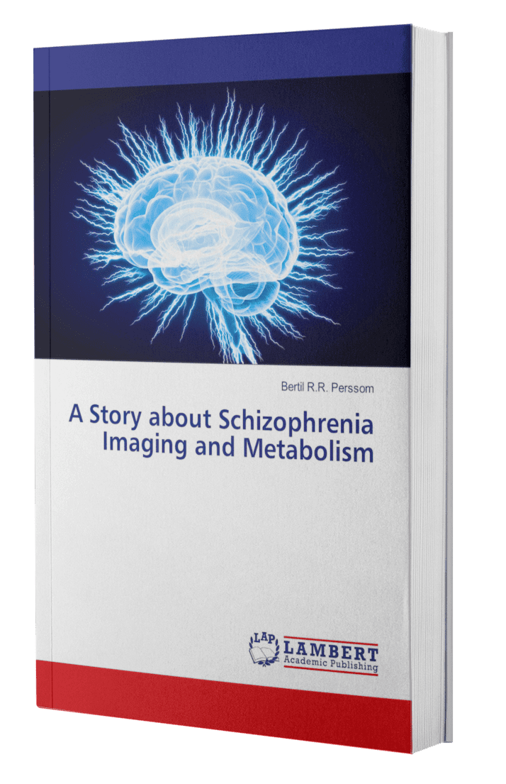
A Story about Schizophrenia Imaging and Metabolism

The author was already in the 1970s involved in Nuclear medicine imaging in Lund where David Ingvar and Göran Franzén studied the brain's regional blood flow in Schizophrenia patients. The introduction of magnetic resonance imaging MRI in the 1980th offered new methods for imaging CSF flow dynamics in the aqueduct. Functional fMRI may be another useful tool for defining the syndrome of Schizophrenia.

Diffusion tensor imaging DTI and its combination with magnetic transfer imaging MTI indicate neuroinflammation in Schizophrenia.MR spectroscopy provides reliable quantification of more than fifteen different brain metabolites such as N-Acetyl Aspartic acid (NAA), Creatine, GABA, Glutamate, and Glutamine in various brain regions. Activation of the Tryptophan metabolism (TRYCAT) pathway appears to be involved in the development and pathophysiology of Schizophrenia.

The dysfunction of the effect of the α-7nicotinic-acetyl-choline receptor (α7nAChR) and/or the N-methyl-D-Aspartate receptor (NMDAR). seems to contribute to cognitive impairment in Schizophrenia motivating new therapeutic strategies The final chapter reviews serum biomarkers.

About the Author

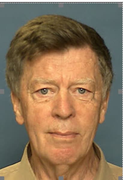
Bertil RR Persson

Rolf Bertil Ragnar PERSSON, PhD, MD.h.c

born : October 12, 1938 in Malmoe, Sweden. 
since 1980-2005 professor of medical radiation physics
2005 –  present: Professor emeritus at Lund University, Sweden

Published : >400 scientific publications,   20  extensive reports and books

Tutor of 40 doctoral dissertations on the faculties of medicine and  science:

After initial studies in the years 1960-62 in chemistry, mathematics and physics, his scientific career began in 1963 with investigations of the health effects of radioactive fallout from the atmospheric nuclear weapons tests, in the lichen-rein-human food chain, which in 1970 resulted in the doctoral thesis: Radioactive fallout in Northern Sweden – Annual variations from 1956 to 1967 in lichens and reindeer and uptake and metabolism in humans.

In the years 1980, 1989-90, 1994 and 1996, he participated in the Swedish polar expeditions to the Arctic (North Pole 10 September 1996) and Antarctica (1989-90) with research in marine radio-ecology. More recently, he has studied the toxicity of naturally occurring radioactive polonium-210 and its distribution in the atmosphere from the North to the South Pole.

His knowledge of chemistry also came in handy in medical radiation physics when the Nordics’ first gamma camera was installed in Lund. This could depict the distribution of radioactive substances in the human body. Previously, they had scanned with a detector, which took a long time and gave poor resolution. But the gamma camera required high levels of radioactive substances in the body to get good pictures. However, it just so happened that an isotope of a new element Teknetium-99m had just been discovered that would be suitable for use with the gamma camera.

His first task was to produce a Technetium-99m sulphur colloid. After diligent experimentation, all the included parameters were optimized and it was time to investigate whether the preparation could be used on patients. The gamma camera images with technetium-99m were a thousand times better than the old scintigraphy with 198Au. This was the beginning of his involvement in medical imaging diagnostics which in 1981 led in unfathomable ways to imaging nuclear magnetic resonance NMR.

Biomedical applications of NMR also involve potential health hazards and he has written extensive monographs on this subject. He has also, in collaboration with the neurosurgeon Leif G Salford, studied the health effects of exposure to electromagnetic fields corresponding to those used in GSM mobile communications. They found increased permeability to blood albumin in the blood-brain barrier in rats.

Their studies of genes in rat brain also show that GSM microwaves do not induce DNA strand breaks or alter chromatin, but rather affect RNA expression under specific exposure conditions.

When in 2015 a national research resource for magnetic resonance imaging MRI came to Lund with a 7-tesla magnet, opportunities opened up to study the brain´s function and chemistry. This stimulated his visions of the chemistry of the soul and also led to his involvement in brain imaging of patients with Schizophrenia which is the subject of this book.

Scroll to Top
Scroll to Top